
Figure 4.
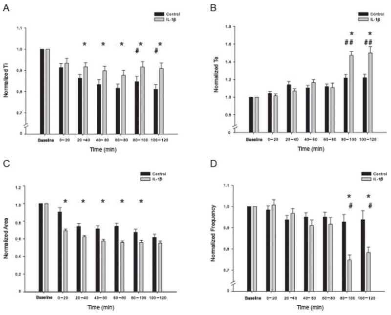
Injection of IL-1β into the caudal/commissural region of the nTS alters fictive inspiratory output from the in vitro en bloc brainstem-spinal cord. (A) Ti remained close to baseline levels after IL-1β injection while control animals saw a time-dependent decrease in activity. (B) Te was significantly higher than control for both time-points after 80 minutes (147.3 ± 4.3, 150.1 ± 6.9%; 121.8 ± 3.9, 121.9 ± 3.8% of baseline respectively). (C) Burst area is significantly smaller after IL-1β injection when compared to control. The burst area is decreased immediately after injection and continues until 100 minutes post injection (see results). The burst area is still smaller after 100 minutes but the difference is no longer statistically significant. (D) Summary data for IL-1β and control burst frequency. Frequency was significantly higher than control for both time-points after 80 minutes (74.7 ± 2.4, 78.2 ± 2.7%; 92.7 ± 3.5, 93.8 ± 4.2 % of baseline respectfully). * indicates significant difference from control group at each time point (p<.05); # indicates significant difference across time (indicated over the control or IL-1β bars).