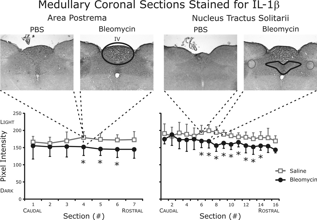
Fig. 8.
Quantification of IL-1β staining in area postrema and nuclei tractus solitarii (nTS). Histological examination identified significant increases in IL-1β in both the area postrema and the nTS. TNF-α and IL-6 staining was similar between the groups in both brainstem areas examined (data not shown).

