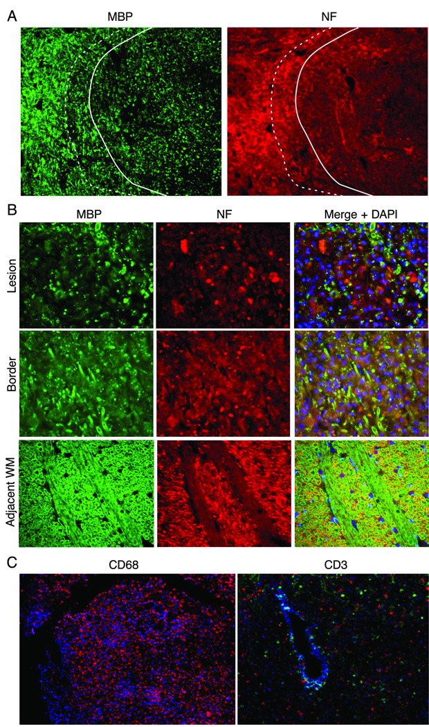
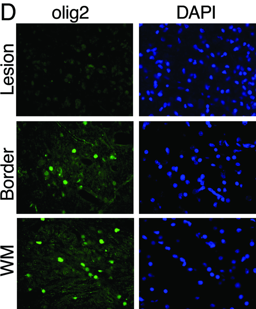
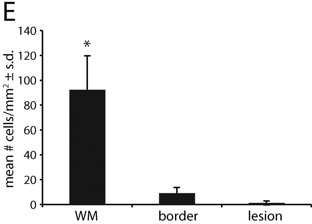
Figure 4. Demyelination and axonal loss in JME lesion.
Immunofluorescent imaging from pontine lesion from JM #13221 identified by MRI (Figure 2J–L) .(A) The pontine lesion was double labeled for MBP (green) and NF (red) to visualize demyelination and loss of neurons. Area to the right of the solid line demarcates demyelinated lesion and shows marked reduction in staining for MBP and NF, indicating loss of myelin and axons; dashed line is lesion border with mix of damaged myelin and normal appearing myelin. Area to the left of the dashed line is unaffected white matter (WM) (5× magnification). (B) High magnification images of the lesion, border and unaffected white matter stained for the presence of MBP, NF and DAPI for nuclei to visualize the extent of demyelination and axonal damage. Note reduction in MBP and NF staining in both the lesion and border areas. The merged image of lesion and border reveals increased cellularity (increased DAPI staining), some preserved myelinated axons and some NF+ axons without myelin. (40× magnification). (C) A cerebellar lesion from JM #13221 showing CD68+ macrophages (red) and CD3+ T cells (green) near a venule (blue) (10× and 20× magnification, respectively). (D) The lesion site in (A) was immunostained for olig2 to assess the presence of oligodendrocytes and with a nuclear stain (DAPI) in the lesion, border and unaffected white matter (WM). Note the reduction in staining in the sections from the lesion and border areas for olig2 with increased numbers of DAPI+ cells (40× magnification). (E) Quantification of olig2 immuno-labeling. Three adjacent sections were analyzed and three random fields in each section were counted at the lesion, lesion border, and in adjacent white matter, showing significant reduction in olig2+ cells in the lesion and border compared with normal white matter (p<0.01).


