
Figure 5.
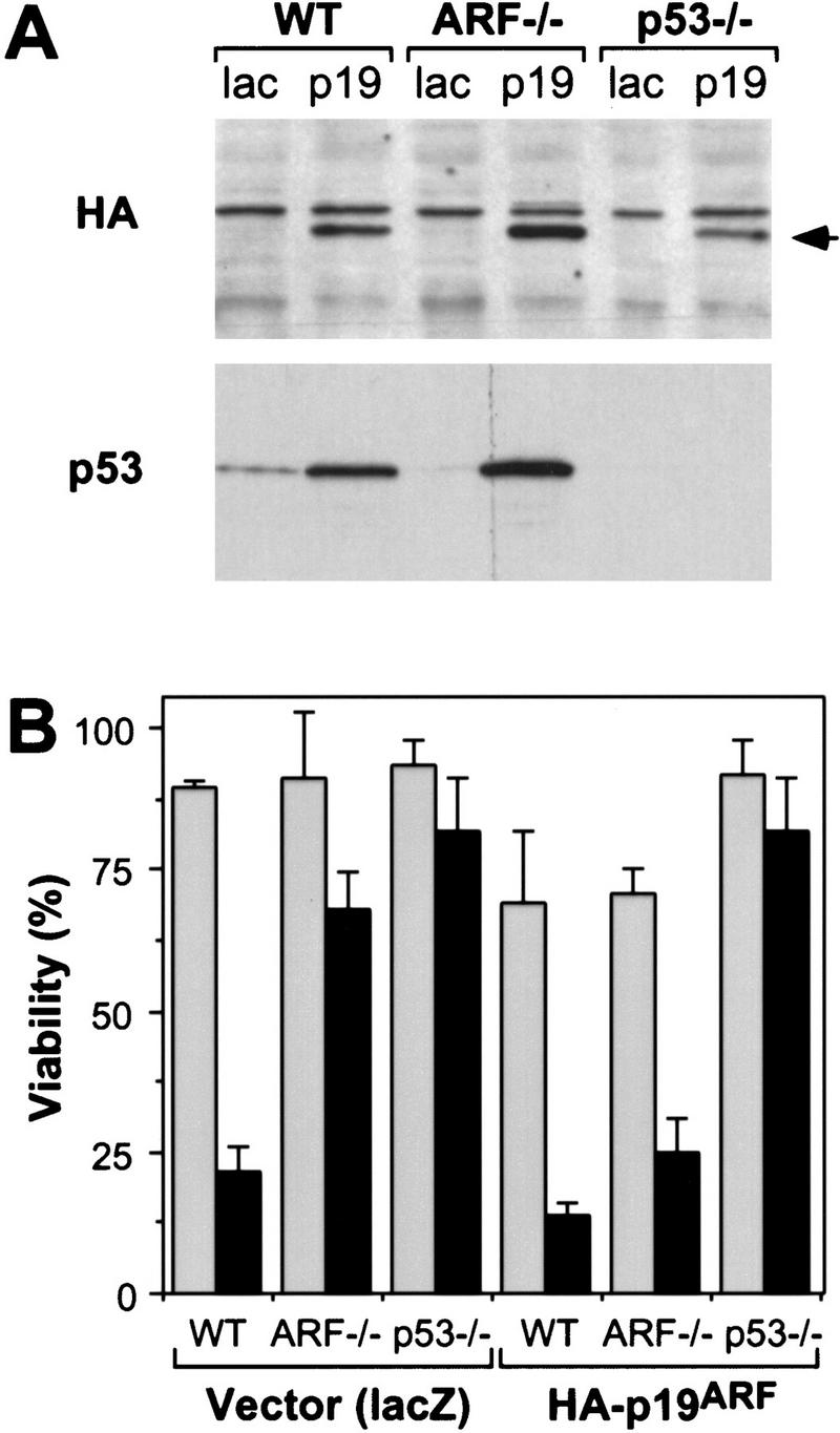
Reintroduction of p19ARF restores apoptosis. Control and E1A-expressing populations derived from wild-type (WT), ARF-null (ARF−/−) and p53-null (p53−/−) populations were infected with retroviruses expressing lacZ or an HA-tagged ARF cDNA (Quelle et al. 1995). Thirty-six hours later, the resulting cell populations were analyzed for p53 and exogenous p19ARF protein expression or treated with apoptotic stimuli. (A) Immunoblotting of infected populations using a monoclonal antibody recognizing the HA epitope fused to p19ARF or a polyclonal antibody directed against p53. The arrow denotes the migration of HA-tagged p19ARF. (B) The indicated cell populations were placed in 10% (shaded bars) or 0.1% (solid bars) serum for 24 hr and cell viability was measured by trypan blue exclusion. The values represent the mean and s.d. of at least three separate infections.