
Fig. 4.
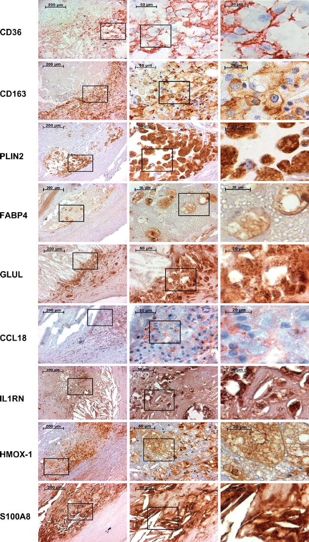
Immunohistochemical stainings of CD36, CD163, PLIN2, FABP4, GLUL, CCL18, IL1RN, HMOX1, and S100A8. The first column displays images obtained from the edge of an atheroma. The second and the third columns are magnifications from foam cell-rich areas indicated by rectangles