Figure 2.

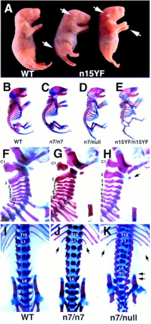
Posterior truncations and homeotic transformations in the Fgfr1 hypomorphs. (A) A newborn wild-type (WT) and two n15YF homozygous (n15YF) pups. Marked reduction in the posterior axis occasionally results in fusion of the hindlimbs (mutant on the right). The mutants also show greatly reduced size of the outer ear, distal limb defects, and severe spina bifida (arrows). Skeletal analysis of a wild type (B), n7 homozygote (C), n7/null transheterozygote (D), and n15YF homozygote (E) demonstrates the series of axial skeletal defects seen in the mutants. Compared to the wild-type cervical vertebrae (F), vertebral malformations are seen with a low frequency in the n7 homozygotes (G). Both the penetrance and expressivity of these defects is increased in the n7/null transheterozygotes (H), which commonly show fusion of the C1 to the occipital bones and partial transformation of C2 into C1 phenotype (arrow). At the lumbrosacral level, compared to the wild type (I), the n7 homozygotes frequently had extra ribs on L1 (J, arrows). Again, these homeotic transformations into the anterior direction were more severe in the n7/null transheterozygotes, which also often showed transformation of S1 into L6 phenotype (K, arrows).
