Figure 7.
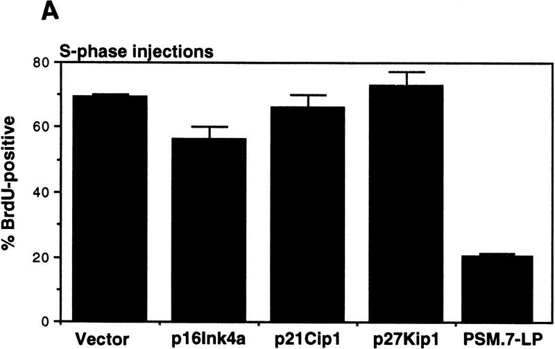
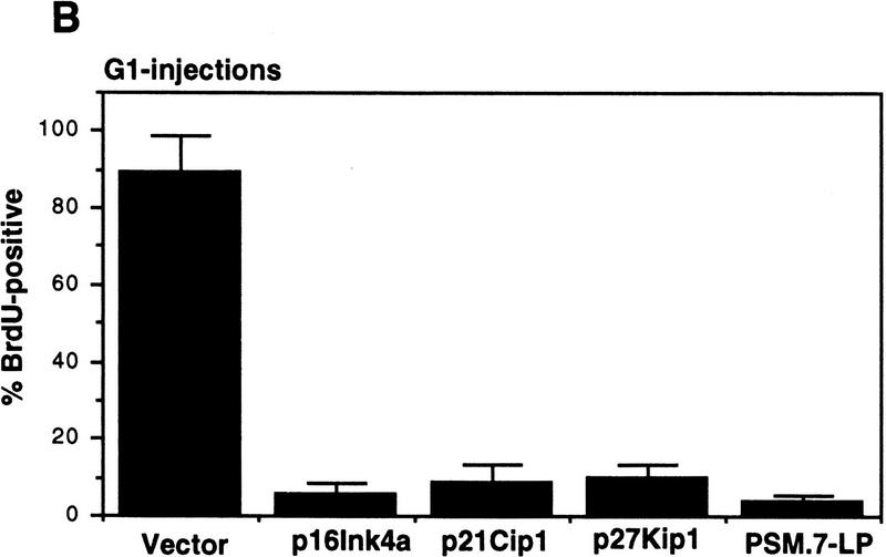
S-phase inhibitory effect of PSM-RB is not observed with cdk inhibitors. (A) Rat-1 cells arrested in S-phase by culturing for 24 hr with aphidicolin were microinjected with the indicated expression plasmids (GFP, 100 ng/μl; p16ink4a, p21cip1, p27kip1, or PSM,7-LP, 50 ng/μl). Following 16 hr more in aphidicolin to allow the accumulation of the plasmid-encoded protein, cells were released from the aphidicolin block and labeled with BrdU for 6 hr. Cells were then fixed and stained for BrdU incorporation. The percentage of GFP-positive cells with BrdU staining was determined. Values shown are the average and deviation from two independent experiments with ∼70 GFP-positive cells counted per experiment. (B) Rat-1 cells were synchronized in early G1 by a 2-hr stimulation of quiescent cells with 10% serum. These early G1 cells were microinjected with the indicated expression plasmids (GFP, 100 ng/μl; p16ink4a, p21cip1, p27kip1, or PSM,7-LP, 50 ng/μl). BrdU was immediately added to the media, cells were labeled continuously for 18 hr and then processed for immunofluorescence analysis. Values shown are the average and deviation from two independent experiments with ∼70 productively injected cells (GFP-positive) counted per experiment.


