Figure 1.

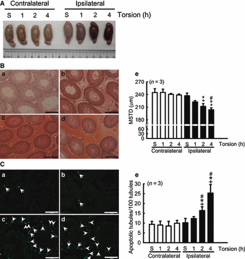
Apparent histological changes and apoptotic cells. (A) After 1 h of torsion, the ipsilateral testis showed congestion and haemorrhage and this was aggravated after 2 and 4 h of testicular ischaemia. (B) Oedema and haemorrhage were observed in the interstitial spaces, after (c) 2 h and (d) 4 h of torsion. Histological sections of the rats from the (a) sham and (b) 1-h groups show normal seminiferous tubular architecture (stain: haematoxylin and eosin; scale bar, 200 μm). (e) The quantifications of mean seminiferous tubular diameter of bilateral testes. (C) Testis sections stained by the terminal deoxynucleotidyl transferase-mediated dUTP-biotin nick-end labelling (TUNEL) method demonstrate the presence of apoptotic cells in testes (a–d). Prominent apoptotic spermatogonia (a–d, arrows) and interstitial cells (c and d, arrow head) in the ipsilateral testes are indicated. (e) Quantitative comparisons of TUNEL-stained cells after different periods of torsion (scale bar, 100 μm). Values are mean ± SEM. *p<0.05, vs. respectively contralateral testes; +p<0.05, vs. sham (S) group; #p<0.05, vs. ipsilateral 1 h group.
