
Figure 4.
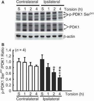
Torsion decreased the phosphorylation of phosphoinositide-dependent protein kinase-1 (PDK1). (A) A representative gel pattern shows the expression of p-PDK1 Ser241 and PDK1 after different periods of torsion. Blotting shows PDK1 (two bands) and p-PDK1 Ser241 (three bands) at 58–68 kDa, and β-actin at 43 kDa. β-actin was used as an internal control. (B) The ratio of p-PDK1 Ser241/PDK1 was decreased after 2 and 4 h of torsion in the ipsilateral testis. *p<0.05, vs. respectively contralateral testes; +p<0.05, vs. sham (S) group; #p<0.05, vs. ipsilateral 1 h group.