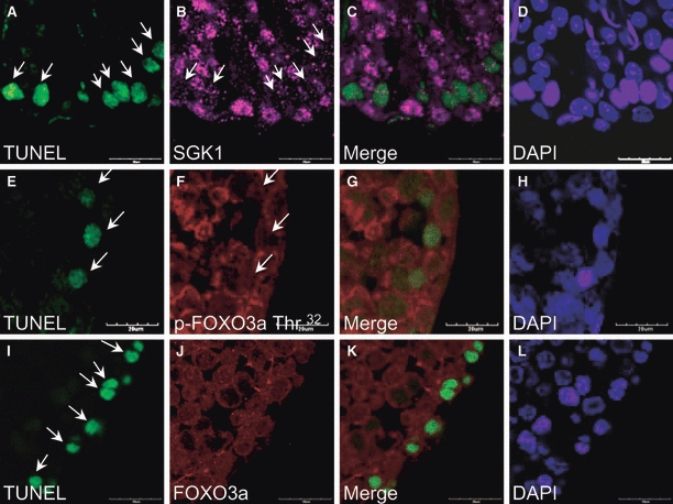
Figure 6.

Immunofluorescent terminal deoxynucleotidyl transferase-mediated dUTP-biotin nick-end labelling (TUNEL) staining of serum- and glucocorticoid-inducible kinase-1 (SGK1)/TUNEL, p-FOXO3a Thr32/TUNEL and FOXO3a/TUNEL in the ipsilateral 4-h testis sections. The apoptosis cells (arrows) are pinpointed by TUNEL stain (green; A, E, and I), whereas the expression of SGK1 (purple; B) and p-FOXO3a Thr32 (red; F) is barely detectable. The expression of FOXO3a (red; J) is not directly relatable to identifiable apoptotic cells. The colocalization images of SGK1/TUNEL, p-FOXO3a Thr32/TUNEL and FOXO3a/TUNEL are shown in C, G and K. The sections are counterstained with DAPI (blue; D, H and I). Scale bar: 20 μm.
