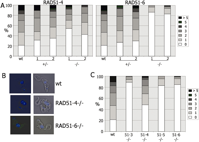
Fig. 4.

RAD51 subnuclear foci formation in T. brucei bloodstream form RAD51 paralogue mutants. A. Quantification of RAD51 foci formation in wild-type (wt) cells compared with two independent heterozygous (+/− 1, 2) or homozygous (−/− 1, 2) mutants of RAD51-4 or RAD51-6 after growth for 18 h in 1.0 µg ml−1 phleomycin; the number of subnuclear RAD51 foci in individual cells is shown as a percentage of the population (counting > 100 cells). B. Examples of RAD51 localization in wt, rad51-4−/−and rad51-6−/− heterozygous cells after 18 h growth in 1.0 µg ml−1 phleomycin. Cells were visualized by differential interference contrast (DIC), DNA was stained with DAPI, and RAD51 was visualized using polyclonal anti-RAD51 antiserum and SFX-conjugated goat-derived anti-rabbit secondary. Merged DAPI and RAD51 (left), and DAPI, RAD51 and DIC (right), images are shown. C. Quantification of RAD51 foci in wt, rad51−/−, rad51-3−/−, rad51-4−/−, rad51-5−/− and rad51-6−/− cells.
