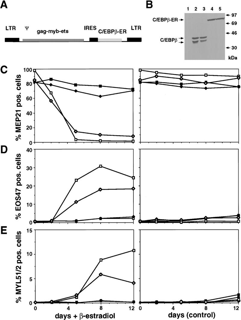
Figure 7.

Effects of activating chicken C/EBPβ in MEPs transformed by E26–βER virus. (A) Structure of the E26–βER virus. (B) Western blot analysis of C/EBPβ–ER protein in E26–βER MEPs (βER cl.1 and cl.2; lanes 4,5) and of endogenous C/EBPβ in E26-WT-transformed myeloblasts (lanes 2,3). An E26–WT control MEP clone is shown for comparison (WT cl.1; lane 1). Western blotting was done with anti-chicken C/EBPβ antiserum on equal protein amounts from each clone. Bands corresponding to the endogenous C/EBPβ and exogenous C/EBPβ–ER are indicated. (C) Down-regulation of MEP21 expression by C/EBPβ–ER activation. E26–WT and E26–βER-transformed MEPs were treated with β-estradiol or mock treated, and their expression of MEP21 antigen, (D) EOS47 antigen, and (E) MYL51/2 antigen measured by FACS analysis after 2, 5, 8, and 12 days. Data from two representative clones of each type are shown. (▪) E26–WTcl.1; (♦) E26–WTcl.2; ( ) E26–βERcl.1; (⋄) E26–βERcl.2.
