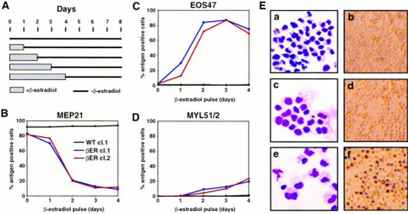
Figure 8.

Effect of transient β-estradiol treatment on E26–βER-transformed MEP clones. (A) Experimental protocol. Cells were treated for 1–4 days with β-estradiol, thoroughly washed, and incubated in the absence of the hormone for a total of 8 days, at which time they were subjected to FACS analysis. Changes in MEP21 (B), EOS47 (C), and MYL51/2 (D) expression in MEP clones transformed by E26–WT and E26–βER after transient exposure to β-estradiol for 1–4 days (as indicated) were determined by antibody staining and FACS analysis at day 8 after initiation of the experiment. (Black line) E26–WTcl.1; (blue line) E26–βERcl.1; (red line) E26–βERcl.2. (E) May–Gruenwald–Giemsa (left) and peroxidase staining (right) of untreated E26–βER cl.1 cells (a,b), or cells treated for 1 day (c,d), or 2 days (e,f) with β-estradiol. Note that 30% of the cells in c and d are EOS47 positive.
