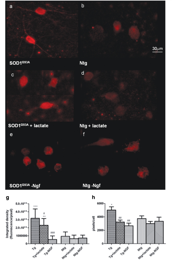
Figure 6.
p75 expression in motoneuron cultured for 14 days on SOD1G93A or Ntg astrocytes under basal conditions (a and b), with 1mM lactate supplementation (c and d) and Ngf depletion (e and f). p75 fluorescence has been quantified and expressed as integrated density (g) in order to capture the quantity of p75 expressed and pixel/cell (h) in order to quantify the area positive for p75. Statistical analysis was performed comparing all groups to p75 expression in motoneurons grown on Ntg (significance =*, where *=<0.05; **= < 0.01; ***= < 0.001) or SOD1G93A astrocytes (significance = # where #= <0.05; ##= < 0.01; ###= < 0.001) under basal conditions (n = 60, bars = SD, one-way ANOVA). Our results show that motoneurons cultured under the influence of SOD1G93A astrocytes show expression of p75 within dendrites and axons as well as the perikarya (a), which is absent in all other conditions (b–f). Consistently, fluorescence quantification (g–h) shows that p75 intensity is significantly higher in normal motoneurons co-cultured for 14 days with SOD1G93A astrocytes compared to motoneurons co-cultured with Ntg astrocytes (P < 0.0001) and the area positive for p75 is significantly higher (P < 0.05). Both lactate supplementation and NGF depletion cause a significant reduction in the area positive for p75, bringing it to levels comparable to those observed in non-transgenic co-cultures. The intensity of p75, nevertheless, reaches normal levels in motoneurons cultured with transgenic astrocytes only after NGF depletion (P < 0.0001).

