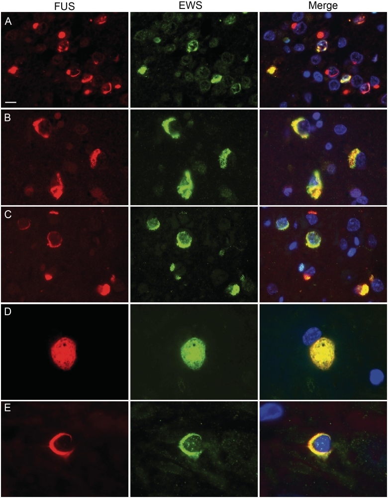
Figure 4.
Co-localization of EWS and FUS in FTLD-FUS inclusions. Double-label immunofluorescence for FUS (red) and EWS (green), with DAPI staining of nuclei in the merged images. In atypical FTLD-U, only a subset of FUS-positive neuronal cytoplasmic and intranuclear inclusions were stained for EWS (A). In contrast, robust co-labelling for EWS and FUS was observed in most inclusions in NIFID (B) and BIBD (C). Inclusions in the lower motor neurons (D, BIBD case) as well as glial cytoplasmic inclusions (E, BIBD case) also showed co-localization. Scale bar: A–C = 10 µm; D = 6.5 µm; E = 4 µm.

