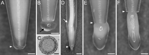
Fig. 3.
Morphology of the root apices in various stages of passing through the narrow zone in variant I. (A, B) Root apex in the tube, (B, arrow) mucilage and loose cap cells outside the tube, (C) cross-section of the treated apex at the root–cap boundary level, (D) buckling of the root body (arrow), (E) bulb-shaped root apex after leaving the tube, (F) root apex far from the tube ending slowly returning to its typical morphology. White arrowheads in A and D–F indicate the tube end. Scale bars: 0.5 mm (A, B, E, F), 0.1 mm (C), 1 mm (D).

