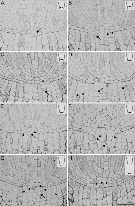
Fig. 6.
Cell pattern at the pole of root apices in variant I seen in median longitudinal sections; the position of the root apex in the tube is shown in the small insets in the upper right corners of the photographs. (A) Three cell layers (arrow) formed at the pole of the root between the vascular cylinder and the cap. (B, C) Meristem opening starts (arrows) by breaking the root–cap boundary, with periclinal divisions (arrowheads) finally leading to cell expansion towards the cap. (D) Atypical longitudinal (arrows) and oblique (arrowheads) cell divisions in columella and the irregular line of the root-cap junction can be seen. (E) Meristem opening due to strong growth of the cell at the pole on the cap side (arrow), with atypical periclinal divisions (arrowheads) in the neighbouring cells. (F) A greatly disturbed root–cap boundary (arrow), with oblique divisions (arrowheads) in the cells protruding on the cap side. (G) The meristem organization is less disturbed, with cells growing through the root–cap boundary on the cap side (arrow), and atypical oblique and longitudinal divisions (arrowheads) both in the root proper and in the cap. (H) Closed meristem, with atypical periclinal divisions (arrowheads) in the middle tier. Scale bar: 50 μm (A–H).

