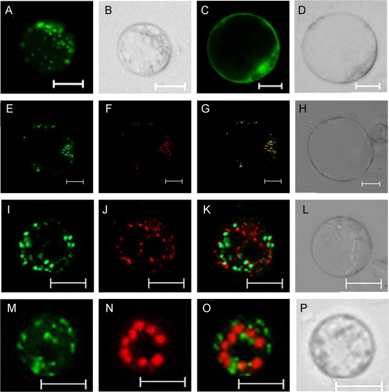
Fig. 2.
Subcellular localization of OsGMST1-GFP transiently expressed in rice protoplasts. (A, B) A rice protoplast cell expressing OsGMST1-GFP (A) and its DIC image (B), showing a punctuate expression pattern. Bars=10 μm. (C, D) A rice protoplast cell expressing GFP (C) as control and its DIC image (D), showing its distribution in nucleus, membrane, and cytoplasm. Bars=10 μm. (E–H) A rice protoplast cell expressing OsGMST1-GFP (E), Golgi localized ST-RFP (F) ,a merged image (G), and its DIC image (H). Bars=10 μm. (I–L) A rice protoplast expressing OsGMST1-GFP (I), stained by mitochondria dye MitoTracker Red (J), a merged image (K), and its DIC image (L). Bars=10 μm. (M–P) A rice protoplast cell expressing OsGMST1-GFP (M), the chlorophyll autofluorescence (N), a merged image (O), and its DIC image (P). Bars=10 μm. (This figure is available in colour at JXB online.)

