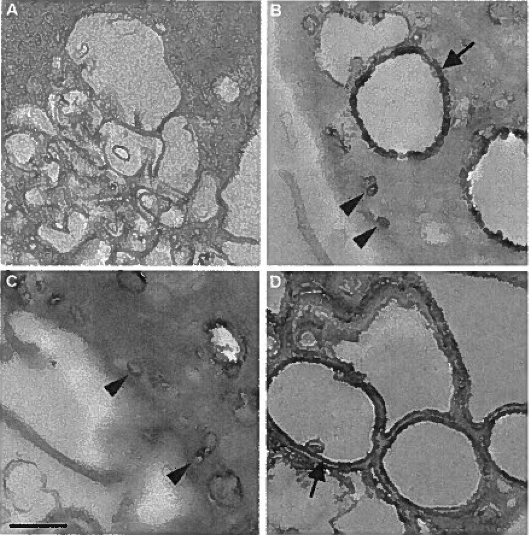
Figure 5.
A through D, Electron microscopy of macrophage uptake of the fluid-phase pinocytosis tracer HRP within macropinosomes and micropinosomes. Macrophages were incubated for 10 minutes without HRP as a control (A) or with 1 mg/mL HRP (B) without inhibitor addition, with 10 µmol/L SU6656 (C), or with 500 nmol/L bafilomycin A1 (D). Arrows and arrowheads indicate macropinosomes and micropinosomes, respectively. The bar indicates 0.5 µm.

