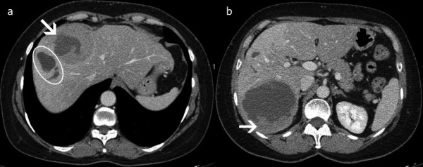
Figure 2.

CT of the liver after chemoembolization. Two partially necrotic lesions remained. The first, 8.9 cm in diameter, extended into segments VI and VII, and the second, 4.2 cm in diameter, extended into segments VIII and IVA. (a) Arrow indicates the necrosis within the lesion. The circle indicates necrosis of the normal liver parenchyma adjacent to the lesion in segment VIII. (b) The viable tumor at the periphery of the lesion is shown.
