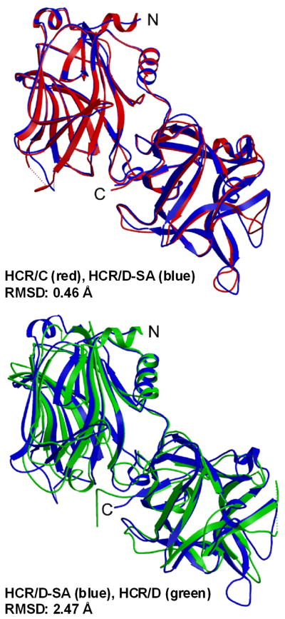
Figure 3. Crystal structures of HCR/C, HCR/D, and HCR/D-SA.
Shown are overlays of the crystal structure of the HCR/D-SA (blue) with HCR/C (left panel, red) RMSD: 0.46 Δ and HCR/D-SA (blue) with HCR/D (right panel, green) RMSD: 2.47Δ. PDB: HCR/C, 3N7K; HCR/D, 3N7J; HCR/D-SA, 3N7L. Reproduced from [62] with permission.

