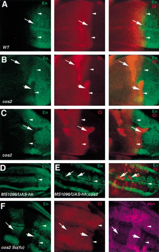
Figure 8.
Cos2 transduces high levels of Hh signaling activity by antagonizing Su(fu). All discs were immunostained with anti-En (green) and/or anti-Ci (red) antibodies. Arrowheads in each panel indicate the A/P compartment boundary. (A) A wild-type wing disc. A compartment cells immediately adjacent to the compartment boundary express En and show low levels of Ci staining (arrow). (B) A wing disc carrying cos2 mutant clones. cos2 mutant cells immediately adjacent to the A/P compartment boundary accumulate high levels of Ci and show diminished levels of En expression (big arrowhead). (C) A clone of cos2 mutant cells derived from the A compartment invade into the P compartment (arrow). The cos2 clone is originated from the A compartment because it expresses Ci but not En. (D) A wing disc expressing MS1096/UAS-hh shows uniform En expression in the wing pouch region of the A compartment. (E) A wing disc expressing MS1096/UAS-hh and carrying cos2 mutant clones. cos2 mutant cells situated in the A compartment accumulate high levels of Ci and show diminished levels of En expression (arrows). (F) A wing disc carrying clones of cos2 Su(fu) double mutant cells and triply stained with anti-En (green), anti-Ci (red) and anti-Myc (purple) antibodies. cos2 Su(fu) double mutant cells are recognized by the lack of Myc expression. cos2 Su(fu) double mutant cells immediately adjacent to the A/P compartment boundary (big arrowhead) or distant from the compartment boundary (arrow) express En and show low levels of Ci. Note that the apparent non-uniform En staining along the dorsoventral axis in D and F is because the mutant wing discs are folded and some of the En expressing cells are out of focus.

