Figure 5.

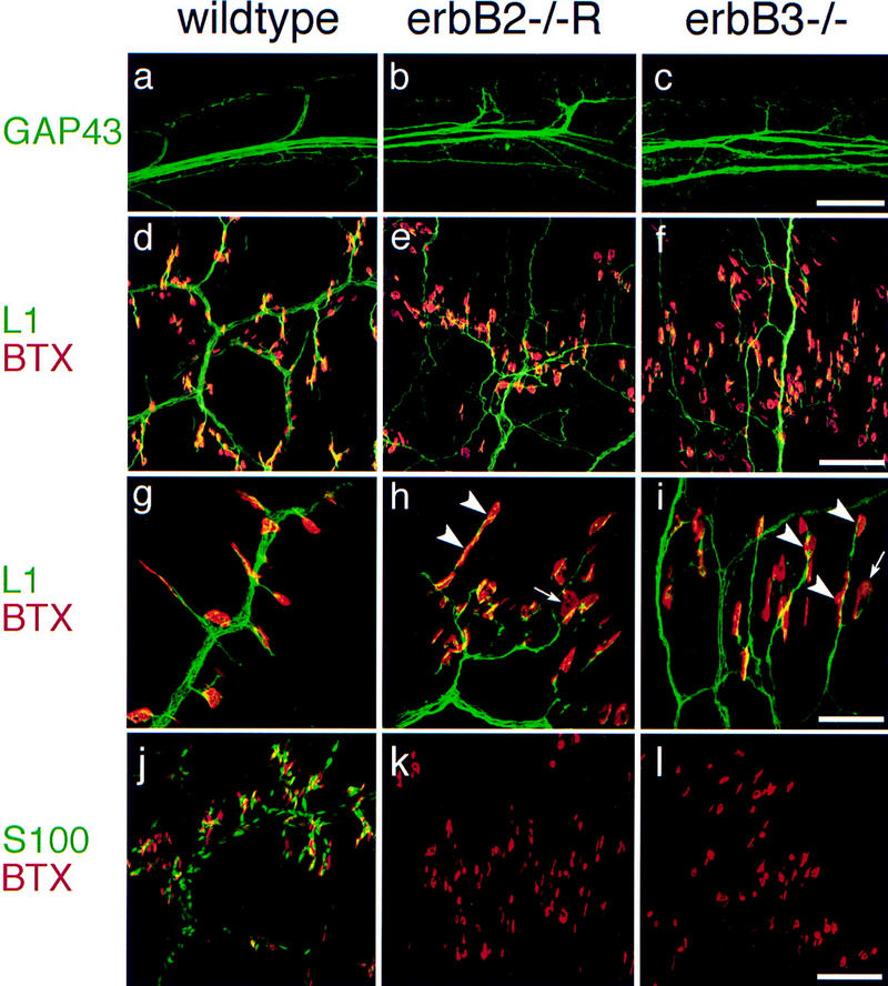
Absence of terminal Schwann cells, morphology of motor nerves, and distribution of neuromuscular synapses in rescued erbB2 mutants and in erbB3 mutants. (a–c) Appearance of proximal nerve branches of 5th intercostal nerves as visualized anti-GAP43 antibodies in wild-type embryos (a), rescued erbB2 (b), and in erbB3 (c) mutants at E15.5. (d–i) Distal projections of thoracic nerves (11th intercostal nerves) and associated synapses at E18.5 (d–f) were visualized with anti-L1 antibodies (green) and rhodamine-labeled α-bungarotoxin (red) in wild-type (d), rescued erbB2 (e), and erbB3 (f) mutant embryos. (g–i) High magnification of synapses associated with distal projections of thoracic nerves in control (g), rescued erbB2 (h), and erbB3 (i) mutant embryos at E18.5; arrowheads point toward multiple synapses associated with one nerve branch, and arrows indicate large AChR clusters. (j–l) Schwann cells and terminal Schwann cells associated with distal projections of thoracic nerves (11th intercostal nerves) and their synapses in control (j), in rescued erbB2 (k), and erbB3 (l) mutant embryos at E18.5. Anti-S100 antibodies (green) and labeled α-bungarotoxin (red) were used to visualize Schwann cells and clustered AChR at the synapse. Bars (a–c), 160 μm; (d–f,j–l) 100 μm; (g–i) 40 μm.
