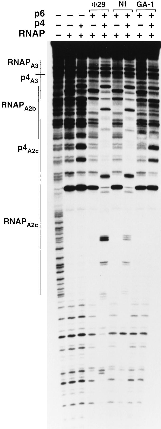
Figure 7.

Complex formation by p6 proteins from related phages in the absence and presence of φ29 p4 protein. The locations of the p4- and RNAP-binding sites are marked with lines. DNase I footprinting reactions included the 366-bp φ29 DNA fragment, B. subtilis ςA–RNA polymerase (20 nm), protein p4 from φ29 (330 nm), and the p6 proteins (14.4 μm) from φ29, Nf, or GA-1, as indicated.
