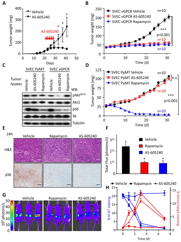
Figure 4. PI3Kγ inhibition halts tumor growth induced by vGPCR.
A, SVEC vGPCR xenografts were prepared as in Figure 1H. Tumors were allowed to grow for 3 weeks (average tumor weight 42.3±2.3 mg) and mice were treated with vehicle or 25 mg/kg AS-605240 twice daily intraperitoneally for five consecutive days. Animal weight was monitored for signs of toxicity. Data points represent mean tumor weight ±SEM (n=8). *, p≤0.001. B, Parallel tumor bearing mice received one treatment with vehicle, rapamycin (5 mg/kg, i.p.), or AS-605240 (25 mg/kg, i.p.) and sacrificed 24 h after. Tumors were excised and processed for H&E staining and pS6 immunohistochemistry. A representative field is shown from 4 tumor samples with similar results. Bar=150μm. C, Tumor xenografts were generated as in panel A using SVEC vGPCR cells expressing the red fluorescent protein mCherry. Tumor growth can be monitored in real time using in vivo fluorescence imaging. Tumors were allowed to grow for three weeks and then treated daily with vehicle, rapamycin (5mg/kg) or AS-605240 (25 mg/kg), intraperitoneally. Photometric analysis of the tumors red fluorescence after one month of treatment shows marked reduction in size in the rapamycin and AS-605240 treated groups. Three representative animals of each group (n=5) are shown. D, Quantification of the tumors from the experiment showed in panel C. Bars represent the average Total Flux (n=10 tumors) ±SEM. E, Tumors generated in parallel as in panel B were excised and processed for immunostainings for the proliferation marker Ki-67 and the apoptotic marker cleaved Caspase3 (Active Casp3) after the indicated days of treatment. Representative areas of every preparation were automatically quantified by software analysis (Supplemental Experimental Procedures). Data points represent the mean percentage ±SEM of positive cells in each preparation. F, Western blot analysis of pAktS473 and pS6 in SVEC PyMT or SVEC vGPCR tumor tissues collected 4h after i.p. injection of the indicated treatments, as described below. G and H, Tumor xenografts of SVEC vGPCR or SVEC PyMT were established as described in Experimental Procedures. Tumors were treated with vehicle, AS-605240 (25 mg/kg/day i.p.) or rapamycin (5 mg/kg/day i.p.), as indicated. Tumor size was measured three times a week. Values represent mean tumor weight ± SEM.

