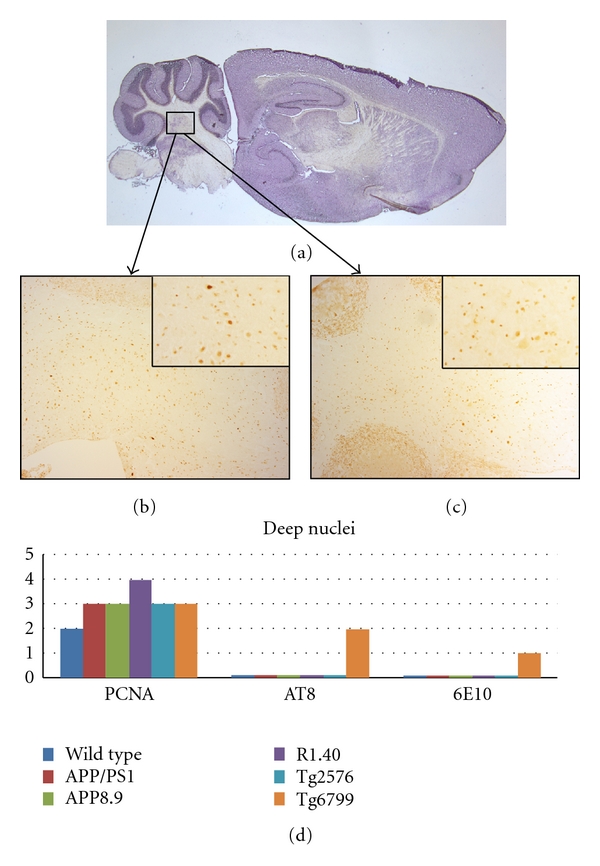
Figure 4.

Comparison among the mouse lines studied with respect to the presence of cell cycle events (PCNA), tau-phosphorylation (AT8), and beta-amyloid plaque deposition (6E10) in the deep cerebellar nuclei. (a) Sagittal section of a wild-type mouse brain indicating the approximate location of the deep nuclei. (b) PCNA-positive neurons are illustrated by their appearance in this representative section from the Tg2576 mouse model. (c) Curiously, some PCNA immunostaining is also seen in wild-type mice as illustrated in this representative micrograph. The insets in both (b) and (c) are representative fields shown at higher magnification to illustrate the qualities of the cell cycle staining. (d) Quantification of the extent of immunostaining for the cell cycle, phospho-tau, and beta-amyloid plaques in the five transgenic models plus wild type.
