Figure 5.

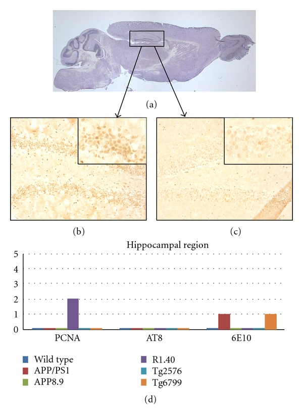
Comparison among the mouse lines studied with respect to the presence of cell cycle events (PCNA), tau-phosphorylation (AT8), and beta-amyloid plaque deposition (6E10) in the hippocampus. (a) Sagittal section of a wild-type mouse brain indicating the approximate location of the areas illustrated in (b) and (c). (b) PCNA-positive neurons are illustrated by their appearance in this representative section from the R1.40 mouse model. Note the involvement of a subset of the dentate granule cells as well as a few CA4 pyramidal neurons (arrows). (c) Minor PCNA immunostaining is seen in wild-type mice. The insets in panels (b) and (c) represent higher magnification of the CA2 region of their respective mouse model. (d) Quantification of the extent of immunostaining for the cell cycle, phospho-tau, and beta-amyloid plaques in the five transgenic models plus wild type. Note that only the R1.40 model showed cell cycle protein expression at the ages we examined.
