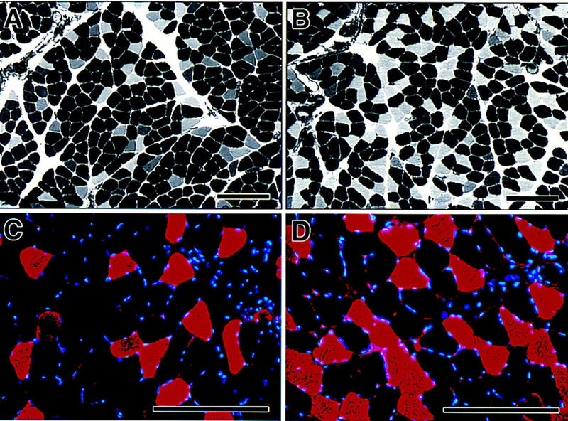
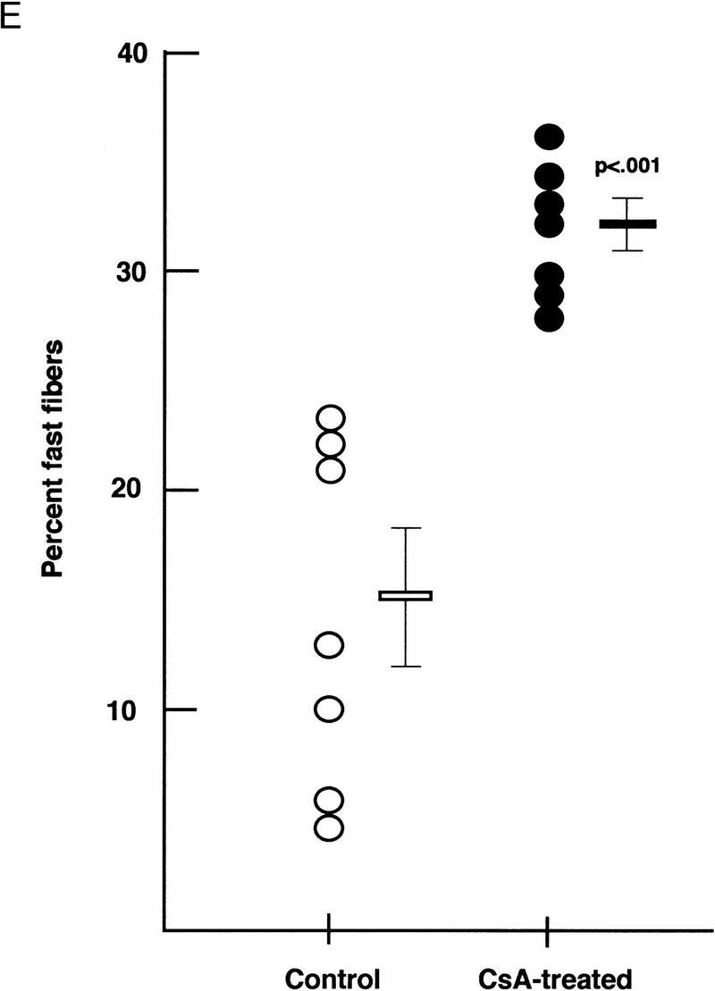
Figure 4.


Fiber composition of soleus muscles from intact rats treated with cyclosporin A. Myosin ATPase activity determined by pH-dependent histochemistry distinguishes slow (darkly stained) and fast (unstained) fibers in sections of soleus muscle from vehicle-treated (A) and cyclosporin A-treated (B) rats. Immunohistochemistry using an antibody raised against fast myosin heavy chain identifies fibers expressing fast myosin (red) in sections of soleus muscle from vehicle-treated (C) and cyclosporin A-treated (D) rats. Nuclei are stained blue. (Bar, 200 μm). (E) Circles represent individual animals. (○) Vehicle treated; (•) cyclosporin A treated and mean values in each group (± s.e.) are shown as horizontal lines. The difference in group means was highly significant (P < 0.001 by unpaired Student’s t-test).
