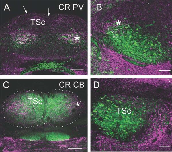
Figure 10.
Fluorescence double labeling of calcium-binding proteins in the torus semicircularis. Transverse sections through caudal (A,C), middle (B), and rostral (D) levels of TSc. Medial is to the right for both B and D. A,B: CR (cyan) and PV: A: In caudal torus, CR labeled terminals in the lateral portion of TSc (compare with CR-ir terminals in Figure 10D). At this very caudal level, most PV-ir neurons overlapped with the CR-ir terminals (asterisk). Arrows mark the pial surface. B: In TSc (left), the ventral PV-ir neuron region extends laterally from the CR-ir terminal recipient region (asterisk) to the ventromedial Tsc (compare with the PV-ir label here and in Fig. 11). Some CR-ir terminals surrounded PV-ir neurons. C: CR and CB in caudal torus (outlined by dots). CR-ir terminals (asterisk) were confined to lateral TSc, whereas CB-ir defined the entire TSc. D: CR and CB in rostral torus. There are few CR-ir terminals in the TSc, but CB-ir defines the extent of TSc. Scale bars = 100 μm in A,C; 200 μm in B,D.

