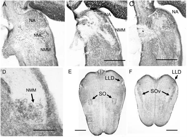
Figure 2.
Calcium-binding protein and SV2 immunoreactivity in horizontal sections. A: NM, NML, and NA are CR-ir and innervated by CR-ir auditory nerve fibers. B,C: SV2 immunoreactivity is absent in the auditory nerve and thus reveals the unlabeled dorsal-caudal fiber bundle (B) bifurcating to the dorsal NMM, lateral NML, and lateral NA. The ventral-rostral fiber bundle (C) projects to medial NA. D: SV2 immunoreactivity in NMM reveals SV2-ir perisomatic terminals. E: CR immunoreactivity in LLD and SO. F: In a more ventral section, CR-ir terminals in lemniscal nuclei and SOv. CR-ir fibers connect the olivary and lemniscal nuclei. Scale bar = 400 μm B (applies to A,B); 400 μm C; 100 μm in D; 600 μm in E,F.

