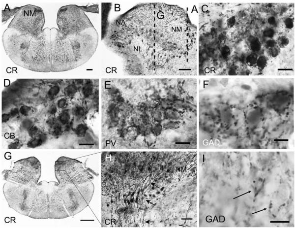
Figure 3.
Calcium-binding protein and GAD immunoreactivity in nucleus magnocellularis and nucleus laminaris. A,C: CR heavily labeled both cell bodies and axonal arbors of NM neurons in transverse sections. B: Sagittal section reveals CR-ir neurons and fibers in NM, NA, and NL; dashed lines indicate the plane of section for A,G. D: PV-ir auditory nerve terminals surround NM neurons. E: CB-ir labels auditory nerve fibers and terminals and also lightly labels NM neurons. F: GAD-ir terminals surround unlabeled somata in NM. G: More rostral section shows NM, NL, and the SO and SOv. H: Enlarged region showing two strands of CR-ir NL bipolar cells ventral to NM; arrows point to dorsal dendrites, cell body, and ventral dendrites. I: GAD-ir terminals surround unlabeled somata in NL. Scale bars = 200 μm in A; 100 μm in B; 20 μm in C–E; 10 μm in F,I; 500 μm in G; 50 μm in H.

