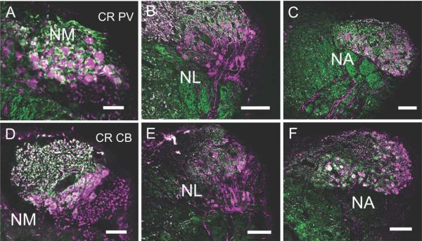
Figure 4.
Fluorescence double labeling of calcium-binding proteins in NM, NL, and NA. The top row shows CR (cyan) and PV (green) immunoreactivity, while the bottom row shows CR and CB (green), all in transverse sections, with medial to the right. A,D: NM. A: CR- and PV-ir labels many axons and terminals around CR-ir NM neurons. Note double labeled (white) auditory nerve terminals. D: CB and CR are co-localized in the auditory nerve terminals and in NM cytoplasm, with CB found in a ring under the cell membrane. B,E: NL. B: PV was absent from NL, although double labeled PV and CR labeled the auditory nerve fibers above NL in NM. CR-ir in NL shows a similar staining pattern as in the HRP immunohistochemistry (Figure 3H). E: CB-ir was present at low levels in NM cytoplasm, but not detected in NL. CR-ir fibers descended to eventually coalesce into the lemniscus, below SO. C,F: NA. C: NA cell bodies are CR-ir, while PV is only localized to terminals around neurons. F: CR heavily labeled many cell bodies in the medial area of NA. Some more lateral neurons expressed both CR and CB, and some neurons express either CR or GB. Note CR-ir fibers descended to join lemniscal fibers. Scale bars = 100 μm.

