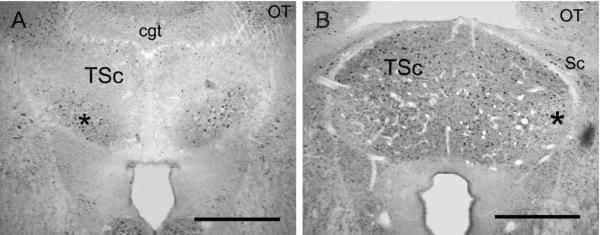
Figure 8.
Calbindin and parvalbumin immunoreactivity in TSc. A: PV-ir cell bodies and neuropil predominate in the ventral TSc (asterisk). B: CB-ir delineates the TSc. Note the decreased CB-ir expression in the lateral region of TSc that receives CR-ir terminals (asterisk). cgt, Commissure of griseum tectale; OT, optic tectum; Sc, preisthmic superficial area. Scale bars = 500 μm.

