
Figure 9.
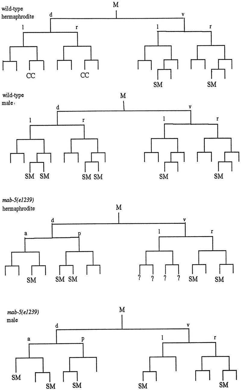
Requirements for MAB-5 in M lineage patterning. Divisions and cell fates in the M lineage are shown for wild-type (Sulston and Horvitz 1979) and sample animals of genotype mab-5(e1239); him-5(e1490) (SM) Sex myoblast; (CC) Coelomocyte; (?) not followed; Cells that apparently adopted a body wall muscle fate are unmarked. To characterize the extent of lineage variability and identify consistent features in the mutant, lineages were followed from the L1 to L3 stage in three hermaphrodites and two males. General features of the mutant lineages are summarized as follows: at hatching, M had migrated toward the posterior (as in wild type) in all mutant animals but was often situated dorsal to wild type. The first division of M was generally normal (although M divided along the anterior–posterior axis in one animal). In the second division, an abnormality was seen in the cleavage plane of Md: this cell frequently divided along the anterior–posterior instead of left–right body axis (n = 5/9 divisions observed in hermaphrodites and 2/3 divisions observed in males). The most striking and consistent phenotypes seen in mab-5(−) hermaphrodites were the loss of the two M-derived coelomocytes (100% of spot-checked animals, with n > 200) and the production of cells that appeared to be supernumary SMs. Like wild-type SMs, these supernumerary myoblasts became enlarged and divided during the L3 stage. The putative SMs arose in multiple, variable positions after M had undergone four divisions (generally without the additional round of cell division that normally occurs in the Mvl/rpa position). The following behavior was seen in the two lineaged hermaphrodites not shown in the figure: The first generated one dorsal SM (Md.ppa) and one ventral SM (Mv.rpa) on the right and two SMs (origins unknown) on the left. All of these cells migrated toward the gonadal anchor cell, enlarged, and underwent multiple cell divisions during the L3. In a second animal in which te Md/vl lineages were followed to mid-L2, the M.vlpa cell became an SM (which migrated toward the gonad) and there were no apparent dorsal SMs. In the additional male lineage, M.drpa and M.vrpa both became apparent SMs that migrated toward the gonad, enlarged, and divided during the L3. To augment this analysis, the early M cell division pattern and the pattern of SMs were observed in larger numbers of animals by spot-checking cell number and position (by Nomarski microscopy) in appropriately staged larvae. Among 27 left or right sides observed in L3 hermaphrodites, 1 side had zero SMs, 7 sides had one SM, 11 sides had two SMs, 6 sides had three SMs, 1 side had four SMs, and 1 side had six SMs. Among 4 left or right sides spot-checked in males, 1 had one SM, 2 had two SMs, and 1 side had four SMs. Generally the resulting cells could be seen in the ventral quadrant, having migrated to flank the center of the gonad; in a minority of cases, the cells apparently migrated to dorsal midbody region (presumably dorsally generated cells that migrated anteriorly but not ventrally) or, less frequently, in more posterior ventral or dorsal positions (presumably SM cells that failed to migrate). The male SMs normally migrate posteriorly, indicating that mab-5 controls the direction of migration of these cells in males. M-derived male mating structures are generally absent in mab-5(0) mutants (see Kenyon 1986).