
Figure 4.
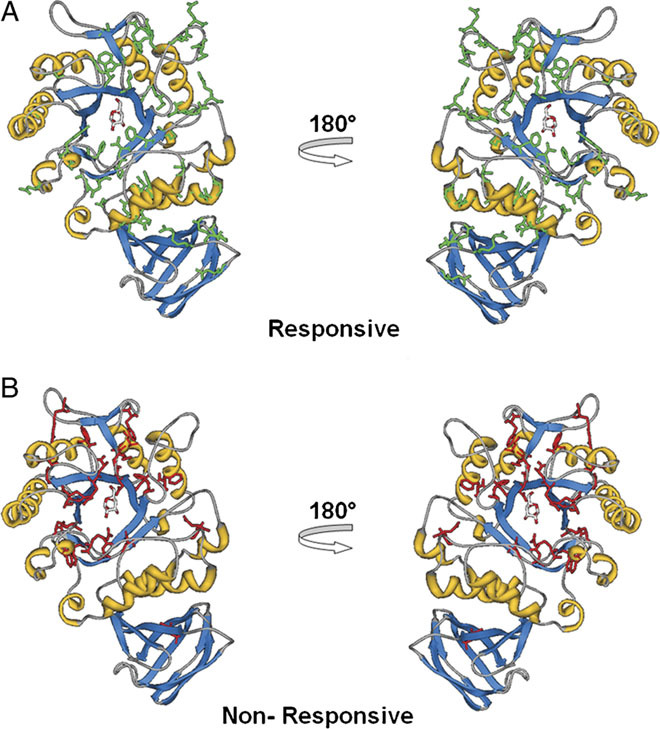
Locations of residues with point mutations in mutant forms of α-Gal A. Eighty-one amino acid residues corresponding to the missense mutations tested in the HEK-293 cell-based assay were mapped onto the structure of the α-Gal A monomer. The α-Gal A monomeric structure is shown in ribbon representation with the bound galactose ligand and affected residues displayed in stick format. Residues with point mutations in AT1001-responsive (A) and nonresponsive (B) mutant forms of α-Gal A are colored green and red, respectively.