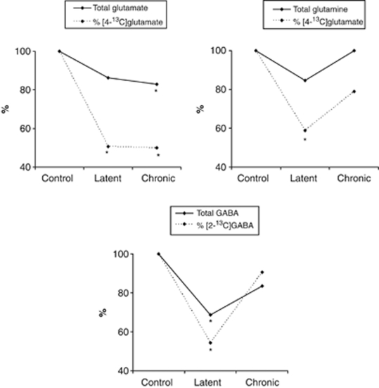
Figure 3.
Glutamate, glutamine, and γ-aminobutyric acid (GABA) in the hippocampal formations of animals in the latent and chronic phase of the kainate (KA) model of mesial temporal lobe epilepsy. Total concentrations (μmol/g tissue) of glutamate, glutamine, and GABA, and their percentage 13C enrichment, as measured in the hippocampal formation by 1H and 13C magnetic resonance spectroscopy (MRS). Values are given for KA-treated animals as percentage relative to controls (100%). The experimental groups comprised animals in the latent phase (2 weeks after KA injection) and animals in the chronic phase (13 weeks after KA injection, calculated from Alvestad et al, 2008), with their respective age-matched saline-treated controls. The [1-13C]glucose was injected 15 minutes before decapitation. For 1H MRS: latent controls n=9, latent KA n=11, chronic controls n=6, and chronic KA n=8. For 13C MRS: latent controls n=8, latent KA n=9, chronic controls n=6, and chronic KA n=8. %, Enrichment signifies turnover of the metabolite, and is expressed as percentage of 13C-label in the fourth position of glutamate and glutamine and in the second position of GABA, as measured by 13C MRS, relative to the total concentration measured by 1H MRS. *Significantly different from control group using Student's t-test, P<0.05.

