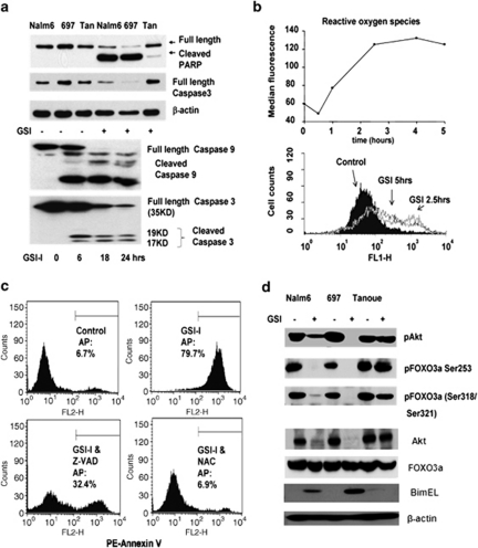
Figure 3.
Induction of proapoptotic pathways after GSI-I treatment. (a) Top: GSI-I treatment leads to PARP and caspase 3 cleavage/activation in Nalm6 and 697 cells but not Tanoue cells. Bottom: time course of caspase 9 and caspase 3 activation in GSI-I-treated 697 cells. (b) Time course of ROS production in 697 cells after addition of 2.5 μ GSI-I. (c) Protection of GSI-induced apoptosis in 697 cells by the caspase inhibitor, Z-VAD or the ROS scavenger, N-acetylcysteine (NAC). (d) Western blotting results show GSI-induced changes in the total AKT, phospho-AKT, phospho-FoxO3a and level of BimEL in lysates from 697, Nalm6 and Tanoue cells.

