Figure 1.
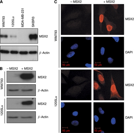
Evaluation of endogenous MSX2 protein expression in WM793 and 1205Lu cells and confirmation of DOX-induced MSX2 over-expression. (A) Western blot analysis of MSX2 protein expression in WM793 and 1205Lu cells. MSX2 migrated at ∼37 kDa. β-Actin levels were used to assess equality of protein loading. The breast cancer cell lines SKBR3 and MDA-MB-231 served as positive and negative controls for the expression of MSX2, respectively. (B) Western blot analysis and (C) immunofluorescent staining for MSX2 in cell lines with conditional MSX2 expression. −MSX2, non-induced cells; +MSX2, DOX-induced cells.

