
Fig. 2.
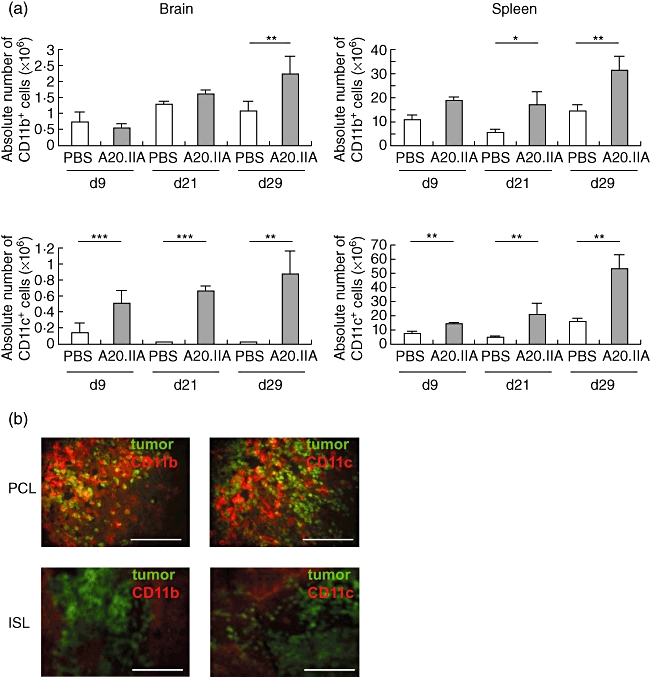
Involvement of antigen-presenting cells in the anti-tumour response in primary cerebral lymphomas (PCL) and intrasplenic lymphoma (ISL). (a) Histograms illustrating the absolute numbers of CD11b+ (upper panels) and of CD11c+ (lower panels) antigen-presenting cells during growth of PCL (left panels) and ISL (right panels), with A20.IIA-green fluorescent protein (GFP) cells, compared with phosphate-buffered saline (PBS)-injected animals, as determined by flow cytometric analyses and trypan blue exclusion (two independent experiments, n = 10). *P < 0·05; **P < 0·005; ***P < 0·0005. (b) Representative photomicrographs of brain and spleen slices at day 21, stained with phycoerythrin (PE)-conjugated anti-CD11b or anti-CD11c antibodies. GFP-positive tumour cells are green. Bars = 200 µm.