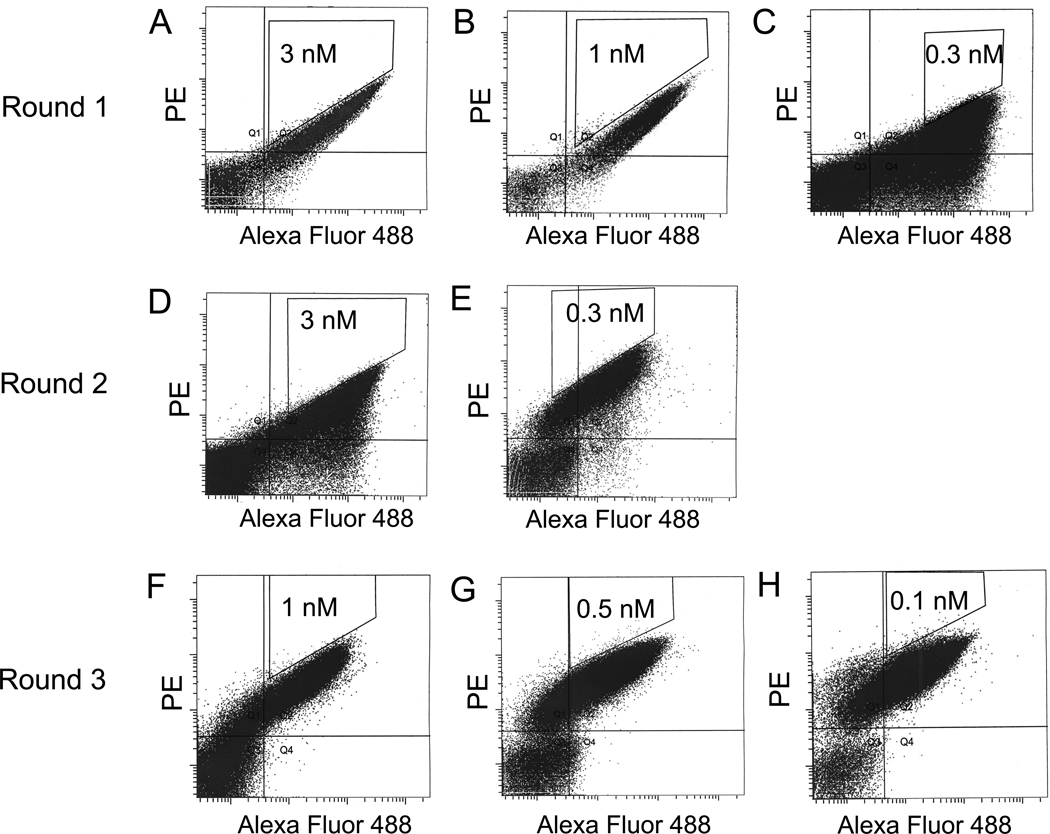
Figure 2. FACS for affinity maturation.
Yeast libraries were labeled with mouse anti-c-myc antibody followed by goat anti-mouse dye as well as biotinylated IGF1 followed by streptavidin-dye. (A, B, C) During three FACS selections of round 1, yeast cells were stained with concentrations of biotinylated IGF1 at 3 nM, 1nM and 0.3 nM, respectively. (D, E) During two FACS selections of round 2, yeast cells were stained with concentrations of biotinylated IGF1 at 3 nM, and 0.3 nM, respectively. (F, G, H) During three FACS selections of round 3, yeast cells were stained with concentrations of biotinylated IGF1 at 1 nM, 0.5nM and 0.1 nM. The 0.1–0.3% cells were selected from sort gates.

