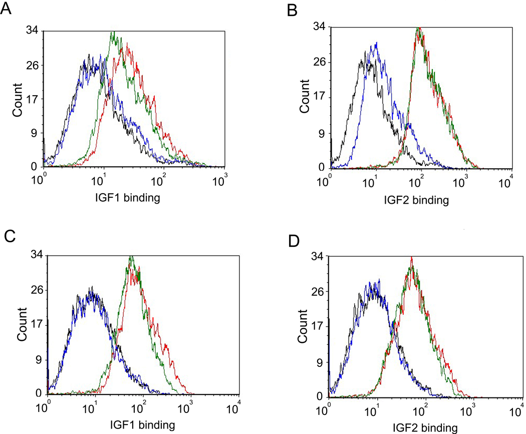
Figure 4. Inhibition of IGF1 and IGF2 binding to MCF-7 cells by m708.5.
(A–B) scFv m708.5 scFv and control scFv 3A2a scFv were pre-incubated with biotinylated IGF1 (A) and biotinylated IGF2 (B) for 20 min at room temperature. Then mixtures were incubated with MCF-7 cells for 30 min on ice. (C–D) IgG1 m708.5 and control IgG1 m102.4 were pre-incubated with biotinylated IGF1 (C) and biotinylated IGF2 (D) for 20 min at room temperature. Then mixtures were incubated with MCF-7 cells for 30 min on ice. After the staining of R-phycoerythrin conjugated streptavidin for 30 min on ice, cells were detected by flow cytometry. In all the graphs, black lines are for cells without any antibody labeling. Blue lines are for tested antibodies, and green lines are for the control antibodies. Red lines show binding with IGF1 or IGF2 alone. Data shown are representatives of three separate experiments performed in duplicates with similar results.

