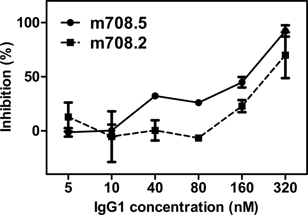
Figure 6. Growth inhibition of MCF-7 cells by m708.2 and m708.5.
MCF-7 cells were incubated in complete medium overnight. Different concentrations of IgG1 m708.2 and m708.5 were pre-incubated with added IGF1 (2.5 nM) and IGF2 (2.5 nM) for 15 min. The media of MCF-7 cells were replaced by the mixture of IgG1 and ligands immediately. Cells were allowed to grow for 3 days, and MTS substrate was added to detect viable cells. The reaction was monitored by absorbance at 450 nM. Positive control was cells in serum free medium with IGF ligands. Blank control was cells in serum free medium without any IGFs. Shown are data with mean ± SEM calculated from three separate experiments.

