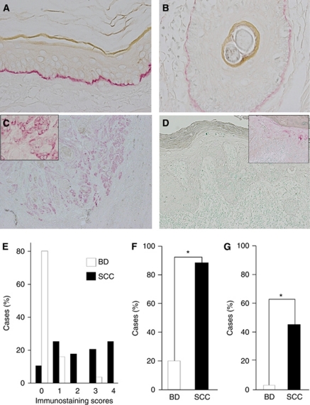
Figure 1.
Immunohistochemical analysis of Ln5-γ2 expression in non-neoplastic skin, squamous cell carcinoma (SCC), and Bowen's disease (BD). (A, B) Non-neoplastic skin showed Ln5-γ2 immunoreactivity along the basement membrane of the epidermis (A) and hair follicle epithelium (B). (C) Squamous cell carcinoma showed Ln5-γ2 immunoreactivity in the cytoplasm of tumour cells, especially in the peripheral portion of tumour nests and the invasion front. (D) Bowen's disease showed only a small number of positive cells within the neoplastic epidermis. (E) Semiquantitative analysis of Ln5-γ2 expression in SCC and BD. (F) Comparison of the proportion of cases positive for Ln5-γ2 expression between SCC vs BD. (G) Comparison of the proportion of cases with high expression (scores 3 and 4) between SCC vs BD. Insets in (C) and (D), magnified views of positive tumour cells. Data are mean±s.e.m. *P<0.01 by Fisher's exact test.

