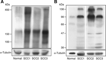
Figure 2.
Detection of Ln5-γ2 and its fragments in tissue extracts from non-neoplastic epidermis and cutaneous SCC by western blotting. (A) Under non-reducing conditions, tissue extracts of the normal epidermis and SCC showed a vague and broad 400–450 kDa band. A 150–180 kDa band was also noted in both tissue extracts, although its intensity was stronger in SCC than normal epidermis. Only SCC tissue showed a band at 100 kDa. (B) Under reducing conditions, both normal skin and SCC tissues showed 130- and 90-kDa bands, although their intensities were stronger in SCC than in normal tissues. Smaller 47- and 30-kDa fragments were also generated in SCC tissue extracts. SCC1-3, three different cases of human SCC.

