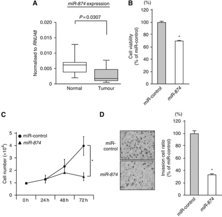
Figure 1.
Expression of miR-874 in MSSCC clinical specimens and gain-of-function study using miR-874 in the IMC-3 cell line. (A) The miR-874 expression levels in clinical specimens. Real-time RT–PCR showed that miRNA expression in tumour tissues was lower than that of normal tissues. RNU48 was used as an internal control. (B) Cell proliferation determined by the XTT assay in the IMC-3 cell line transfected with 10 nM of miR-874 or miR-control. (C) Cell number was counted after transfection with 10 nM of miR-874 or miR-control at 24, 48, and 72 h. (D) Cell invasion activity determined by the Matrigel invasion assay in IMC-3 cell lines transfected with 10 nM of miR-874 or miR-control. *P<0.05.

