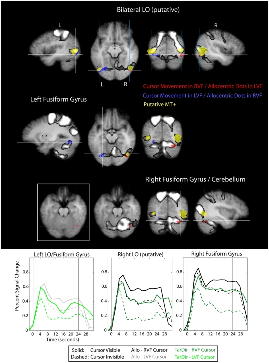
Figure 8.
Results of the conjunction “Allocentric > Target-Directed (Cursor movement in LVF, relevant target-dots in RVF)” and “Allocentric > Target-Directed (Cursor movement in RVF, relevant target-dots in LVF)” (Table 1) with regard to the occipital lobe, overlaid on subjects’ average Talairach-brain (n = 14). Statistical thresholds (p < 0.005) were those applied to each of the component contrasts in the conjunction. Probability of type-I errors in the resulting activation map was controlled using cluster size thresholds. It is evident that activity in the occipital lobe related to allocentric spatial coding included posterior portions of the fusiform gyrus (FFG) and portions of lateral occipital cortex adjacent and posterior to putative visual motion area MT+ (putative area LO). For stereotactic coordinates of activation foci see Table 3. Brain areas in the occipital lobe that are more active during allocentric movements performed in the right or left visual field are denoted in red and blue, respectively. Also shown are “visual feedback areas” (yellow) and the “shared sensory–motor network” (gray). The inset in Figure 8 shows activity without the “shared sensory–motor network” to illustrate the border between cerebellum and activity in inferior occipital cortex. Event related averages of regions depicted in red and blue are shown as well.

