Figure 5.
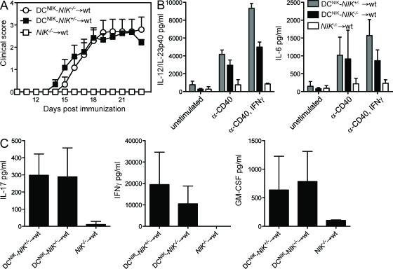
Expression of NIK in DCs is sufficient to restore EAE susceptibility in NIK−/− mice. (A) DCNIK-NIK−/− → WT, DCNIK-NIK+/− → WT, and NIK−/− → WT BMCs were immunized with MOG35–55/CFA and observed for clinical signs of EAE. Shown is one representative of three independent experiments (n = 6). Error bars indicate SEM. (B) Splenic DCs were isolated from DCNIK-NIK−/− → WT and DCNIK-NIK+/− → WT BMCs and stimulated in vitro with α-CD40 and IFN-γ for 24 h. IL-12/IL-23p40 and IL-6 secretion was measured by ELISA. (C) Splenocytes of MOG35–55/CFA-immunized DCNIK-NIK−/− → WT and DCNIK-NIK+/− → WT BMCs were isolated at the peak of disease and restimulated in vitro with MOG35–55 for 48 h. Supernatants were analyzed by ELISA. (B and C) Each graph shows one representative of three independent experiments. Error bars indicate SD.

