Figure 3.
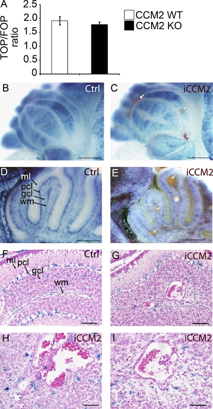
Ccm2 deletion has no significant effect on Wnt–β-catenin signaling pathway in CCM2 KO ECs in vitro and in iCCM2 lesions. (A) TCF/LEF-β-catenin transcriptional activity in CCM2 WT and null ECs in vitro was determined by transfecting CCM2 WT and KO ECs with the TOP-TK-Luc or the FOP-TK-Luc reporter constructs (containing WT or mutant Tcf/Lef binding sites, respectively, and a basal TK promoter, upstream a luciferase gene). Columns are means ± SD of triplicates from a representative experiment out of three performed. (B-I) Animals were bred with the BAT-Gal reporter mouse to assess β-catenin activation (see Materials and methods for breeding details). All animals were injected with tamoxifen at P1. XGal staining, performed on control and iCCM2 cerebellum, is shown (n = 8 in each group, from 3 different litters). White arrows show the CCM lesions in iCCM2 animals (in C, E, and G). In F–I, H&E staining was performed on cerebellum sections, after XGal staining. The box in G is magnified in I. H shows a CCM lesion composed of multiple juxtaposed caverns. Gcl, granular cell layer; ml, molecular layer; pcl, Purkinje cell layer; wm, white matter. Bars: 1 mm (B and C); 500 µm (D and E); 100 µm (F and G); 50 µm (H and I).

