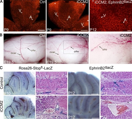
Figure 4.
CCM lesions are capillary-venous and do not affect the arterial compartment. (A) Analysis at P9 or P12 of the retinal vasculature from control or iCCM2 animals using vascular isolectin-B4 staining (left and middle, n = 25 in each group analyzed between P8 and P10), after XGal staining (right, n = 4 in each group, from 2 different litters). Tamoxifen-induced Ccm2 deletion was performed at P1. Arteries (A) are thin and normal in iCCM2 retinas, whereas veins (V) are dilated in iCCM2 animals. Asterisks show CCM lesions developing at the periphery of the retina. (B) Lateral view of cerebral hemispheres of control and iCCM2 embryos dissected at E19.5 (n = 8 in each group, from 4 different litters). Ccm2 deletion was performed at E14.5. Vascular anomalies affect the caudal rhinal vein (crhv) and the capillaries surrounding in the iCCM2 animals. The box in the middle panel is magnified in the image on the right. (C) Analysis of the cerebellar vessels at P10 and P12 after XGal staining on whole brain (left) and after a H&E staining (right). Animals were crossed with either the Rosa26-Stopfl-LacZ reporter mouse or the artery-specific EphrinB2tlacZ reporter mouse. Results shown are representative of at least four animals in each group, from two different litters. Bars: 1 mm (A and B, left and middle); 500 µm (B [right] C [left]); 100 µm (A, right); 50 µm (C, right).

