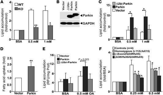
Figure 4. Parkin modulation of lipid uptake requires an intact ubiquitin-like domain.
(A) Lipid accumulation assayed by Nile red staining in MEFs from Parkin+/+ (WT) and Parkin–/– (KO) after BSA-conjugated oleate (0.5 and 1 mM) incubation. Values represent the fold relative to BSA incubation and normalized to cell number. The values represent the average of 5 independent experiments. (B) Parkin expression in HepG2 cells overexpressing vector, WT Parkin, and ∆Ubi-Parkin constructs. (C) Lipid accumulation in HepG2 cells overexpressing vector, Parkin, and ∆Ubi-Parkin after oleate incubation (0.5 and 1 mM). (D) Relative fluorescence of Bodipy-labeled dodecanoic acid at the end of 1,200-second incubation in HepG2 cells overexpressing Parkin compared with vector controls. Values represent the fold relative to vector-transfected HepG2 cells. (E) Lipid accumulation in SH-SY5Y neuroblastoma cells overexpressing vector, Parkin, and ΔUbi-Parkin after 0.5 mM oleate incubation. Values are normalized to protein concentration. (F) Lipid accumulation in response to 0.25 and 0.5 mM oleate in transformed B cells from 3 patients with PARK2 mutations versus 4 WT control subjects. Values were normalized to cell number. Data are displayed relative to control cells exposed to BSA normalized to 1. Data are expressed as mean ± SD. *P < 0.05; **P < 0.01 versus control or vector.

